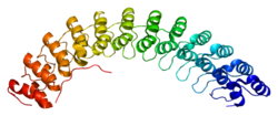
ANK1
Ankirin 1, koji je poznat kao ANK1, je protein koji je kod ljudi kod ljudi kodiran ANK1 genom.[1][2]
| Ankyrin 1, eritrocitski | |||||||||||
|---|---|---|---|---|---|---|---|---|---|---|---|
 PDB grafika bazirana na 1n11. | |||||||||||
| Dostupne strukture | |||||||||||
| 1n11 | |||||||||||
| Identifikatori | |||||||||||
| Simboli | ANK1; ANK; SPH1; SPH2 | ||||||||||
| Vanjski ID | OMIM: 182900 MGI: 88024 HomoloGene: 55427 GeneCards: ANK1 Gene | ||||||||||
| |||||||||||
| Pregled RNK izražavanja | |||||||||||
 | |||||||||||
 | |||||||||||
 | |||||||||||
| podaci | |||||||||||
| Ortolozi | |||||||||||
| Vrsta | Čovek | Miš | |||||||||
| Entrez | 286 | 11733 | |||||||||
| Ensembl | ENSG00000029534 | ENSMUSG00000031543 | |||||||||
| UniProt | P16157 | Q0VGY9 | |||||||||
| RefSeq (mRNA) | NM_000037 | XM_981917 | |||||||||
| RefSeq (protein) | NP_000028 | XP_987011 | |||||||||
| Lokacija (UCSC) |
Chr 8: 41.63 - 41.87 Mb |
Chr 8: 24.44 - 24.62 Mb | |||||||||
| PubMed pretraga | [1] | [2] | |||||||||
Distribucija u tkivu
уредиProtein kodiran ankirin 1 genom je prototip ankirinske familije. On je prvo otkriven u eritrocitima, ali je od tog vremena isto tako pronađen u mozgu i mišićima.[2]
Genetika
уредиKompleksni struktura alternativnog spajanja u regulatornom domenu, stvara različite izoforme ankirina 1, međutim precizna funkcija tih brojnih izoformi nije poznata. Alternativna poliadenilacija koja dovodi do niza eritrocitnih ankirin 1 iRNK molekula različitih veličina je primećena. Skraćene izoforme ankirina 1 specifične za mišiće, koje su proizvod primene jednog alternativnog promotora, su isto tako bile identifikovane.[2]
Povezanost bolesti
уредиMutacije eritrocitnog ankirina 1 su bile asocirane u skoro polovini svih pacijenata sa naslednom sferocitozom.[2]
Funkcija
уредиANK1 protein pripada ankirinskoj familiji koja povezuje integralne membranske proteine sa potpornim spektrin-aktin citoskeletonom, i igra ključnu ulogu u aktivnostima kao što su ćelijska pokretljivost, aktiviranje, proliferacija, kontakt i održavanje specijalizovanih membranskih domena. Više izoformi ankirina sa različitim afinitetima za brojne proteine su izraženi u tkivno-specifičnom razvojno-regulisanom maniru. Većina ankirina se tipično sastoji of tri strukturna domena: amino-terminalni domen koji sadrži višestruka ankirin ponavljanja; centralin region sa visoko konzervisanim spektrin vezivajućim domenom; i karboksi-terminal regulatorni domen koji je najmanje konzerviran i koji je subjekat varijacije.[2]
sAnk1 (engl. small ANK1) varijanta proteinskog spajanja uspostavlja kontakte sa obskurin proteinom, gigantskim proteinom koji okružuje kontraktilni aparat u izbrazdanom mišiću.[3]
Interakcije
уредиANK1 pokazan da ima protein-protein interakciju sa Tiam1 proteinom[4] koji promoviše propagaciju Rac1 signala i tim putem razvoj raka dojke. ANK1 isto tako uspostavlja interakcije sa nizom drugih proteina, kao što su: Titin,[5] RHAG[6] i OBSCN.[7]
Vidi još
уредиReference
уреди- ^ Lambert S; Yu H; Prchal JT (1990). „cDNA sequence for human erythrocyte ankyrin”. Proc. Natl. Acad. Sci. U.S.A. 87 (5): 1730—4. PMC 53556 . PMID 1689849. doi:10.1073/pnas.87.5.1730.
- ^ а б в г д „Entrez Gene: ANK1 ankyrin 1, erythrocytic”.
- ^ Borzok MA, Catino DH, Nicholson JD, Kontrogianni-Konstantopoulos A, Bloch RJ (2007). „Mapping the binding site on small ankyrin 1 for obscurin”. J. Biol. Chem. 282 (44): 32384—96. PMID 17720975. doi:10.1074/jbc.M704089200.
- ^ Bourguignon LY, Zhu H, Shao L, Chen YW (2000). „Ankyrin-Tiam1 interaction promotes Rac1 signaling and metastatic breast tumor cell invasion and migration”. J. Cell Biol. UNITED STATES. 150 (1): 177—91. ISSN 0021-9525. PMID 10893266.
- ^ Kontrogianni-Konstantopoulos, Aikaterini; Bloch Robert J (2003). „The hydrophilic domain of small ankyrin-1 interacts with the two N-terminal immunoglobulin domains of titin”. J. Biol. Chem. United States. 278 (6): 3985—91. ISSN 0021-9258. PMID 12444090. doi:10.1074/jbc.M209012200.
- ^ Nicolas, Virginie; Le Van Kim Caroline; Gane Pierre; Birkenmeier Connie; Cartron Jean-Pierre; Colin Yves; Mouro-Chanteloup Isabelle (2003). „Rh-RhAG/ankyrin-R, a new interaction site between the membrane bilayer and the red cell skeleton, is impaired by Rh(null)-associated mutation”. J. Biol. Chem. United States. 278 (28): 25526—33. ISSN 0021-9258. PMID 12719424. doi:10.1074/jbc.M302816200.
- ^ Kontrogianni-Konstantopoulos, Aikaterini; Jones Ellene M; Van Rossum Damian B; Bloch Robert J (2003). „Obscurin is a ligand for small ankyrin 1 in skeletal muscle”. Mol. Biol. Cell. United States. 14 (3): 1138—48. ISSN 1059-1524. PMID 12631729. doi:10.1091/mbc.E02-07-0411.
Literatura
уреди- Bennett V, Baines AJ (2001). „Spectrin and ankyrin-based pathways: metazoan inventions for integrating cells into tissues.”. Physiol. Rev. 81 (3): 1353—92. PMID 11427698.
- Bennett V (1979). „Immunoreactive forms of human erythrocyte ankyrin are present in diverse cells and tissues.”. Nature. 281 (5732): 597—9. PMID 492324. doi:10.1038/281597a0.
- Lambert S; Yu H; Prchal JT (1990). „cDNA sequence for human erythrocyte ankyrin.”. Proc. Natl. Acad. Sci. U.S.A. 87 (5): 1730—4. PMID 1689849. doi:10.1073/pnas.87.5.1730.
- Fujimoto T, Lee K, Miwa S, Ogawa K (1991). „Immunocytochemical localization of fodrin and ankyrin in bovine chromaffin cells in vitro.”. J. Histochem. Cytochem. 39 (11): 1485—93. PMID 1833445.
- Lux SE, John KM, Bennett V (1990). „Analysis of cDNA for human erythrocyte ankyrin indicates a repeated structure with homology to tissue-differentiation and cell-cycle control proteins.”. Nature. 344 (6261): 36—42. PMID 2137557. doi:10.1038/344036a0.
- Davis LH, Bennett V (1990). „Mapping the binding sites of human erythrocyte ankyrin for the anion exchanger and spectrin.”. J. Biol. Chem. 265 (18): 10589—96. PMID 2141335.
- Korsgren C, Cohen CM (1988). „Associations of human erythrocyte band 4.2. Binding to ankyrin and to the cytoplasmic domain of band 3.”. J. Biol. Chem. 263 (21): 10212—8. PMID 2968981.
- Cianci CD, Giorgi M, Morrow JS (1988). „Phosphorylation of ankyrin down-regulates its cooperative interaction with spectrin and protein 3.”. J. Cell. Biochem. 37 (3): 301—15. PMID 2970468. doi:10.1002/jcb.240370305.
- Steiner JP, Bennett V (1988). „Ankyrin-independent membrane protein-binding sites for brain and erythrocyte spectrin.”. J. Biol. Chem. 263 (28): 14417—25. PMID 2971657.
- Hargreaves WR, Giedd KN, Verkleij A, Branton D (1981). „Reassociation of ankyrin with band 3 in erythrocyte membranes and in lipid vesicles.”. J. Biol. Chem. 255 (24): 11965—72. PMID 6449514.
- Bourguignon LY, Lokeshwar VB, Chen X, Kerrick WG (1994). „Hyaluronic acid-induced lymphocyte signal transduction and HA receptor (GP85/CD44)-cytoskeleton interaction.”. J. Immunol. 151 (12): 6634—44. PMID 7505012.
- Maruyama K, Sugano S (1994). „Oligo-capping: a simple method to replace the cap structure of eukaryotic mRNAs with oligoribonucleotides.”. Gene. 138 (1-2): 171—4. PMID 8125298. doi:10.1016/0378-1119(94)90802-8.
- Morgans CW, Kopito RR (1993). „Association of the brain anion exchanger, AE3, with the repeat domain of ankyrin.”. J. Cell. Sci. 105 (Pt 4): 1137—42. PMID 8227202.
- Bourguignon LY; Jin H; Iida N (1993). „The involvement of ankyrin in the regulation of inositol 1,4,5-trisphosphate receptor-mediated internal Ca2+ release from Ca2+ storage vesicles in mouse T-lymphoma cells.”. J. Biol. Chem. 268 (10): 7290—7. PMID 8385102.
- Eber SW; Gonzalez JM; Lux ML (1996). „Ankyrin-1 mutations are a major cause of dominant and recessive hereditary spherocytosis.”. Nat. Genet. 13 (2): 214—8. PMID 8640229. doi:10.1038/ng0696-214.
- Lanfranchi G; Muraro T; Caldara F (1996). „Identification of 4370 expressed sequence tags from a 3'-end-specific cDNA library of human skeletal muscle by DNA sequencing and filter hybridization.”. Genome Res. 6 (1): 35—42. PMID 8681137. doi:10.1101/gr.6.1.35.
- del Giudice EM; Hayette S; Bozon M (1996). „Ankyrin Napoli: a de novo deletional frameshift mutation in exon 16 of ankyrin gene (ANK1) associated with spherocytosis.”. Br. J. Haematol. 93 (4): 828—34. PMID 8703812. doi:10.1046/j.1365-2141.1996.d01-1746.x.
- Zhou D; Birkenmeier CS; Williams MW (1997). „Small, membrane-bound, alternatively spliced forms of ankyrin 1 associated with the sarcoplasmic reticulum of mammalian skeletal muscle.”. J. Cell Biol. 136 (3): 621—31. PMID 9024692. doi:10.1083/jcb.136.3.621.
- Gallagher PG; Tse WT; Scarpa AL (1997). „Structure and organization of the human ankyrin-1 gene. Basis for complexity of pre-mRNA processing.”. J. Biol. Chem. 272 (31): 19220—8. PMID 9235914. doi:10.1074/jbc.272.31.19220.
- Suzuki Y; Yoshitomo-Nakagawa K; Maruyama K (1997). „Construction and characterization of a full length-enriched and a 5'-end-enriched cDNA library.”. Gene. 200 (1-2): 149—56. PMID 9373149. doi:10.1016/S0378-1119(97)00411-3.
Spoljašnje veze
уреди- ANK1+protein,+human на US National Library of Medicine Medical Subject Headings (MeSH)